
A team of researchers at the California Institute of Technology, led by Professor Changhuei Yang, have figured out a way to crank their microscopy up to 11. Usually, scientists are forced between a rock and a hard place: they can have high res images of small areas or low resolution pictures of larger fields. Using a strategy known as Fourier ptychographic microscopy, Yang's team was able to computationally correct a standard microscope's low res imagery, producing a billion-pixel picture. By adding an LED array to an existing microscope -- the only hardware tweak their $200 system calls for -- the researchers were able to stitch together a 20X quality image from a 2X optical lens. The information gleaned from the LED lights was corrected entirely on a computer, making it an exceptionally cost effective way to create high res microscopic images. The team's report, published by the journal Nature Phototonics, can be read in full at the source link below.
Filed under: Science, Alt
Comments
Via: California Institute of Technology
Source: Nature Phototonics
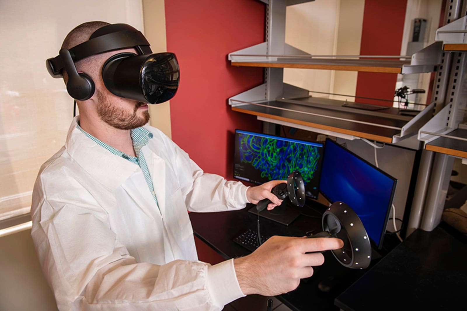 You can only learn so much about cells by studying 2D pictures, and 3D microscope technology can produce an abundance of data that might be hard to decipher. Researchers at Carnegie Mellon University and Virginia Mason have an answer, though: let sci...
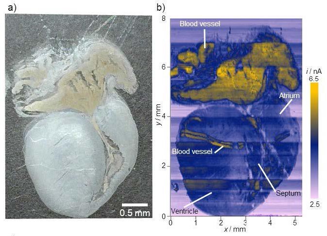 In a study published recently in Angewandte Chemie, researchers demonstrated that an imaging technique called scanning electrochemical microscopy could become a very useful medical tool. Rather than having to use additional chemicals like dyes or flu...


 To date, biologists have typically had to study the progress of a virus through indirect means, such as studying the antibodies -- actually tracking the viruses themselves has been difficult. However, researchers say they've found a way to follow th...
 A collaboration of scientists from University of Washington (UW), the Pasteur Institute and the University of Utrecht have harnessed a state-of-the-art microscope and supercomputer to model a coronavirus' infection mechanism for the first time.

