Holographic tech could be key to future quantum computers

A breakthrough in studying light might just be the ticket to the future of quantum computing. Researchers at EPFL have found a way to determine how light behaves beyond the limitations of wavelengths, opening the door to encoding quantum data in a s...
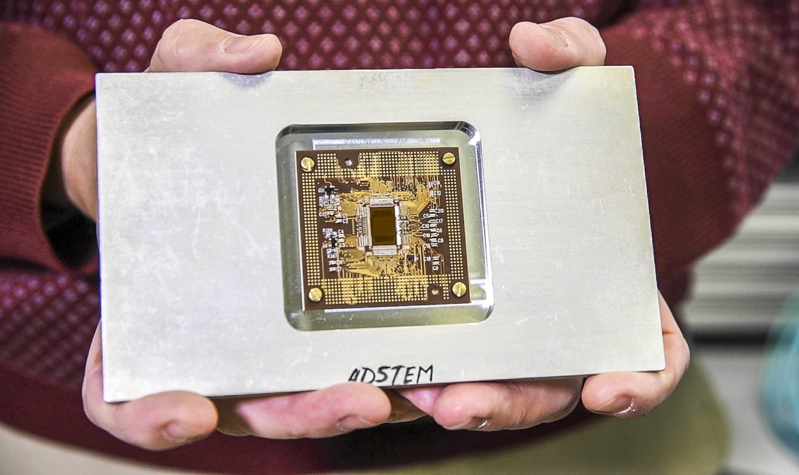
Scientists generate 281-gigapixel cell map using electron microscope

Scientists generate 281gigapixel cell map using electron microscope

Electron microscopes can produce incredibly detailed and even 3D views of sub-cellular structures, but often at the cost of losing the bigger picture. Researchers at Leiden University in the Netherlands, however, have leveraged a technique called virtual nanoscopy that enables researchers to observe the whole of a cell and its intricate details in a single image. With the method, the team stitches together nanometer resolution photographs of what's gone under the scope to create a map with adjustable zoom a la Google Maps. Their study created a 281-gigapixel image (packed with 16 million pixels per inch) of a 1.5-millimeter-long zebrafish embryo. If you'd like to take a gander at the ultra-high resolution fish or read up on the group's findings for yourself, check out the source links below.

Filed under:

Scientists generate 281-gigapixel cell map using electron microscope originally appeared on Engadget on Wed, 08 Aug 2012 04:32:00 EDT. Please see our terms for use of feeds.

Permalink 
  • Via:PhysOrg
  •  | 
  • Source:The Journal of Cell Biology, (2)
  •  | Email this | Comments

    Scientists generate 281-gigapixel cell map using electron microscope

    Scientists generate 281gigapixel cell map using electron microscope

    Electron microscopes can produce incredibly detailed and even 3D views of sub-cellular structures, but often at the cost of losing the bigger picture. Researchers at Liedel University in the Netherlands, however, have leveraged a technique called virtual nanoscopy that enables researchers to observe the whole of a cell and its intricate details in a single image. With the method, the team stitches together nanometer resolution photographs of what's gone under the scope to create a map with adjustable zoom a la Google Maps. Their study created a 281-gigapixel image (packed with 16 million pixels per inch) of a 1.5-millimeter-long zebrafish embryo. If you'd like to take a gander at the ultra-high resolution fish or read up on the group's findings for yourself, check out the source links below.

    Filed under:

    Scientists generate 281-gigapixel cell map using electron microscope originally appeared on Engadget on Wed, 08 Aug 2012 04:32:00 EDT. Please see our terms for use of feeds.

    Permalink PhysOrg  |  sourceThe Journal of Cell Biology, (2)  | Email this | Comments

    Beam-switching endows electron microscopes with 3D, added gross-out

    3D-scanning-electron-microscope-Japanese-research

    Having haunted our curtailed childhoods with tiny, disgusting horrors, the scanning electron microscope is about to get a new lease of life in 3D. Researchers in Japan have figured out how to deflect the electron beam rapidly to give two slightly shifted views, so real-time 3D images can now been scoped on a monitor without even the need for eye-wear. Current gear can only muster flat images, so it's always been painfully slow for scientists to extract convexity and other details from objects. Though the 3D-version is lower-res than the old way, at least now all those slimy mandibles and egg sacs will be right there in your face. Nice.

    Beam-switching endows electron microscopes with 3D, added gross-out originally appeared on Engadget on Thu, 03 May 2012 17:59:00 EDT. Please see our terms for use of feeds.

    Permalink The Verge  |  sourceTechOn!  | Email this | Comments